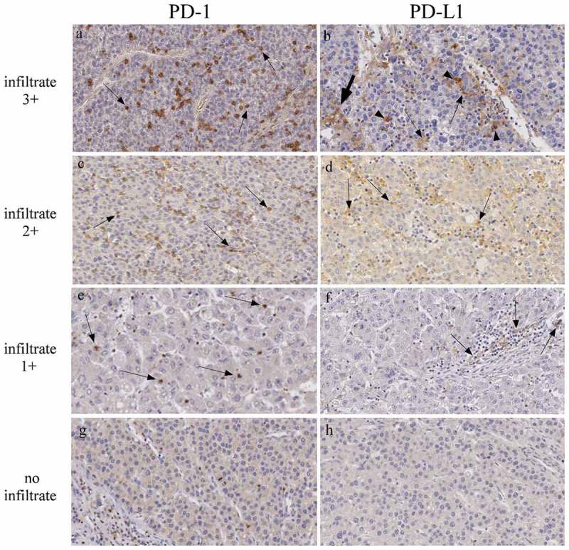
Figure 6.

PD-1 and PD-L1 expression in HCC tumor tissues.
Immunohistochemical staining for PD-1 and PD-L1 in formalin-fixed paraffin-embedded sections of HCC. The left and right panels show representative HCC tumor tissues with very large (infiltrate 3+), large (infiltrate 2+), low (infiltrate 1+), or absent (no infiltrate), lympho-monocyte infiltration (original magnification, x200). Representative of infiltrating PD-1-positive lymphocytes, largely detectable in panel a,c and e, are indicated with arrows. Similarly, representative PD-L1 positive infiltrating cells are indicated by arrows (panels b, d, f). Arrowheads and bold arrow (panel b) indicate liver resident cells, most likely Kupffer cells and hepatic stellate cells, respectively.
