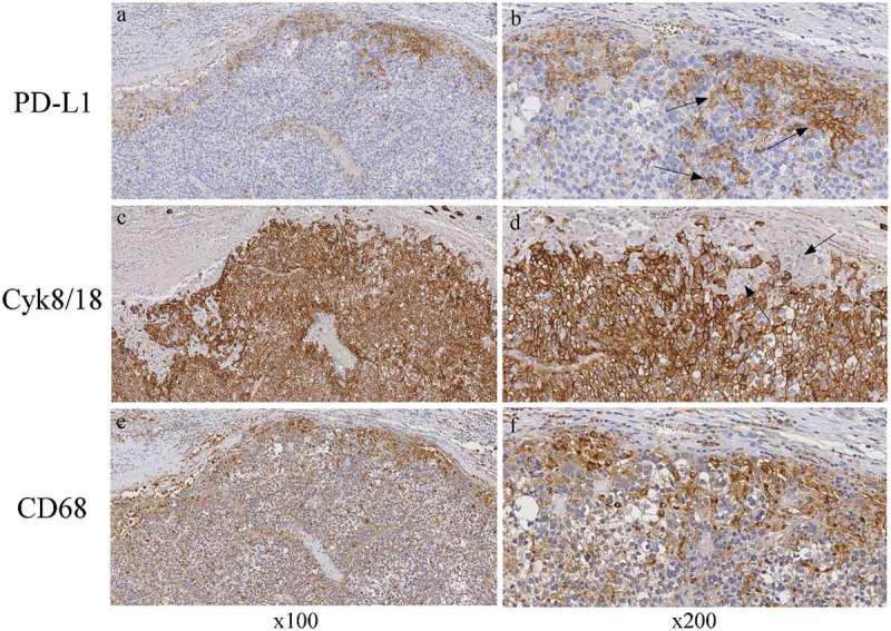
Figure 7.

CD68-positive cells but not Cyk8/18-positive HCC tumor cells co-express PD-L1.
Immunohistochemical staining for PD-L1, CyK8/18 and CD68-positive cells. Photographs showing representative paraffin-embedded blocks of HCC tumor tissue samples with large numbers of CD68-positive myeloid cells concentrated at the margin of the tumor and high PD-L1 expression in the same area (right bottom and top panels, respectively). CyK8/18-positive tumor cells (large brown area in panels c, d) do not overlap with PD-L1-positive cells (panel d, arrows). Left panels original magnification, x100; right panels, original magnification x200.
