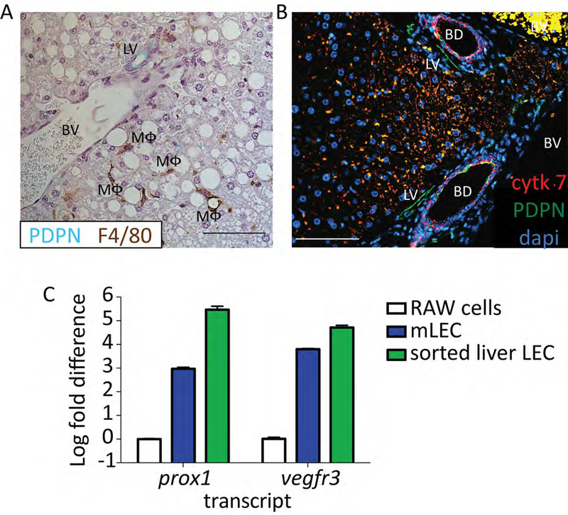
Figure 3.
A. Representative immunohistochemistry from a mouse liver stained with PDPN (blue/green) and F4/80 (brown). Lymphatic vessel (LV), blood vessel (BV) and macrophage (MΦ) are labeled. Scale bar is 100μm. Formalin Fixed paraffin embedded tissue was deparaffinized for 20 minutes in xylene. Hydrated to water through gradient of ethanol, and antigen retrieval was performed using perkin elmer pH6 antigen retrieval buffer in a pressure cooker for 15 minutes. Tissue was blocked using 0.1%BSA and stained using anti-mouse PDPN (8.1.1) 1:400 and anti-mouse F4/80 (MCA497) 1:100 for 1 hour at room temperature. Anti-hamster IgG HRP and anti-rabbit IgG HRP were used as secondary antibodies. DAB+ and Vina Green were used to detect the F4/80 and PDPN respectively. Tissue was counter stained with Hematoxylin and imaged on a Nikon eclipse Ti. B. As in A except PDPN in green, cytokeratin 7 in red and dapi in blue. Tissue was blocked using 5% donkey and 5% goat serum and stained using anti-mouse PDPN (8.1.1) 1:100 and anti-mouse cytokeratin 7 (ab181598) 1:200 for 1 hour at room temperature. Anti-hamster IgG AF647 and anti-rabbit IgG PE were used as secondary antibodies. Tissue was counter stained with DAPI and imaged. Scale bar is 100μm. Lymphatic vessel (LV), blood vessel (BV) and bile duct (BD) are labeled. C. Log fold change in Vegfr3 and prox-1 expression from sorted liver LECs based on staining in protocol (CD45-, CD31+, CD146lo/neg and PDPN+) compared to RAW cells or primary murine lymph node LECs. Sorted cells were passed through a QIAshredder column and RNA was extracted using a QIAgen RNeasy micro kit and cDNA was made using the Qiagen QuantiTect reverse transcription kit. Transcript abundance was normalized to the housekeeping gene, Gapdh for every sample. Primers for Vegfr3, prox-1 and Gapdh were obtained from Qiagen.

