Figure 5.
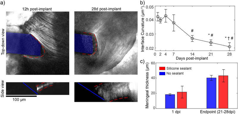
Meningeal remodeling over the first month post implant. (a) Top-down views (top) of meningeal collagen I from the same animal at 12h and 28 days post implant reveals that, collagen-I (outlined in red) remodels to conform to the electrode surface (highlighted in blue). Side views (bottom) show that the meninges thicken (meninges outlined in red) (b) The curvature of collagen-I at the meningeal-electrode interface reduces over the first month, indicating a flatter, more conformal collagen interface (one-way ANOVA p < 0.001; significant reducMons relaMve to 1 dpi (*), 2 dpi (†), and 4 dpi (#) assessed with Tukey’s HSD post hoc tests; p <0.05). This analysis used all silicone sealed animals with visible meningeal-electrode interfaces (n = 4 for 0.5–21dpi, n = 3 for 28dpi) (c) Automated quantification of meningeal thickness within 1 dpi and at the experimental endpoint (between 21–28 dpi) shows statistically significant change in meningeal thickness over time (two-way ANOVA p< 0.005 for time effect; n = 6 for silicone at 1dpi, n = 5 for all other groups)). Sealant condition did not affect meningeal thickness, suggesting that the thickening was not in response to the presence of silicone. All data presented as mean ± SEM.

