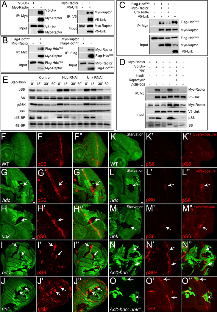
Figure 5. Hdc and Unk Form a Protein Complex with Raptor and Regulate S6 Phosphorylation.
Physical interaction between Unk and Raptor. Immunoprecipitates of S2R+ cell lysate expressing the indicated combination of V5-Unk and Myc-Raptor constructs were probed with the indicated antibodies.
Physical interaction between Hdc70kDa and Raptor. Immunoprecipitates of S2R+ cell lysate expressing the indicated combination of Flag-Hdc70kDa and Myc-Raptor constructs were probed with the indicated antibodies.
Unk potentiates Hdc70kDa-Raptor interaction. S2R+ cells expressing the indicated constructs were analyzed by co-immunoprecipitation (co-IP).
The interaction between Unk and Raptor is nutrient sensitive. S2R+ cells expressing the indicated constructs were analyzed by co-IP.
RNAi of Hdc or Unk induces S6 phosphorylation. S2R+ cells were incubated with double-stranded RNA (dsRNA) of GFP, hdc, or unk for 3 days and treated with PBS starvation for indicated time before western blot. Total cell lysates were probed with anti-phospho-S6, anti-phospho-S6K (T389), and anti-phospho-4E-BP1 (T37/46) antibodies.
(F–F’’) A wild-type control eye disc was stained with pS6 antibody (red).
(G–J’’) Eye discs or wing discs containing mutant clones of the indicated genotypes were stained with pS6 antibody (red). In all panels, mutant clones were marked by loss of GFP. Note increased pS6 staining in hdc or unk mutant clones (arrows).
(K–M’’) Same as (F)–(H’’), except all eye discs were treated with 15 min PBS starvation prior to fixation.
(N–N’’) An eye disc containing hdc-overexpressing clones (GFP-positive) was stained with pS6 antibody (red).
(O–O’’) An eye disc containing unk mutant clones overexpressing hdc (GFP-positive) was stained with pS6 antibody (red).
Images (F), (G’), and (H’) and images (K’), (L’), and (M’) were acquired using the same parameter settings. The scale bars represent 20 μm.

