Figure 5.
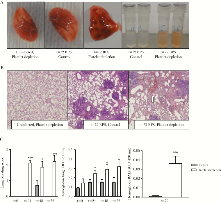
Thrombocytopenia results in lung bleeding at the site of infection. Mice were treated with anti-GPIbα (platelet depletion) or immunoglobulin G (IgG) control (both 0.4 µg/g) and infected with Burkholderia pseudomallei via the airway and killed after 24, 48, or 72 hours or killed uninfected. A, Representative photographs of naive or infected lungs and bronchoalveolar lavage fluid (BALF). B, Representative microphotographs of hematoxylin and eosin (H&E)–stained tissue sections (original magnification ×40), bleeding indicated by arrow. C, Quantification of lung bleeding; both scored on H&E-stained tissue sections by a pathologist blinded for groups and hemoglobin measurement in 50-fold diluted lung homogenates or BALF. Data are represented as bars (mean with standard error of the mean). n = 8 mice per group. *P < .05, ***P < .001 vs IgG control. Abbreviations: BALF, bronchoalveolar lavage fluid; BPS, B. pseudomallei; OD, optical density.

