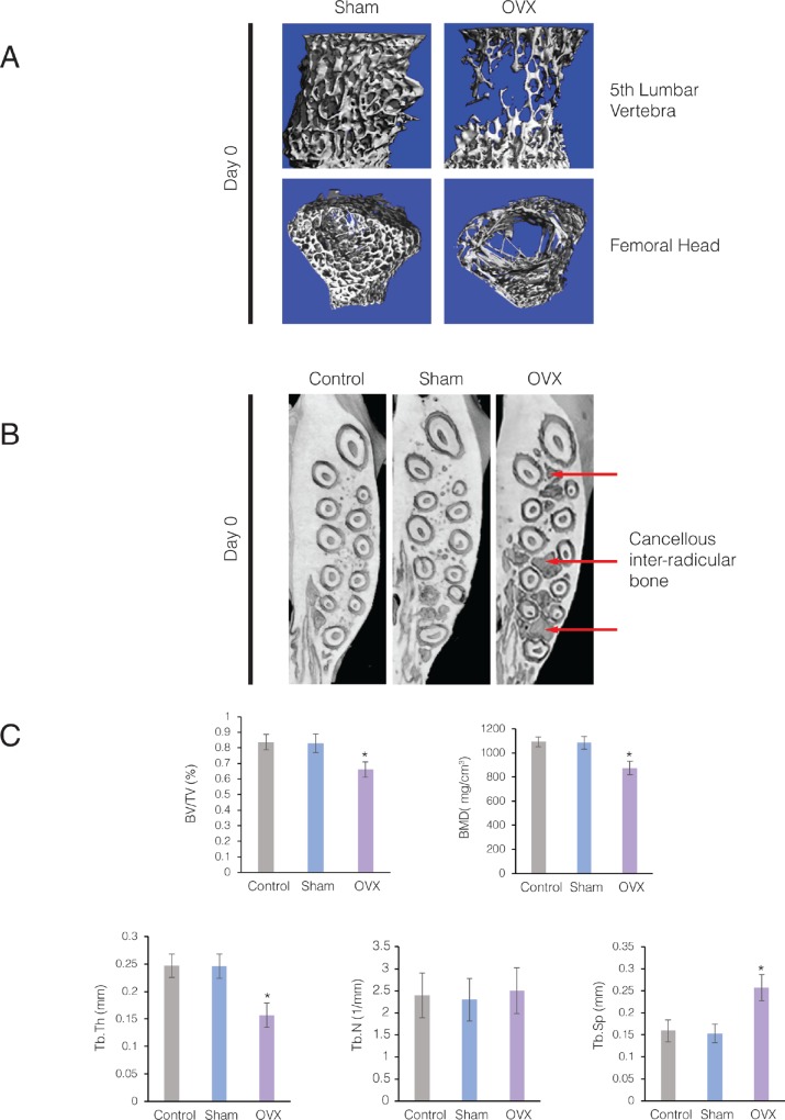
Fig 4. Ovariectomized rats on low mineral diet developed osteoporotic alveolar bone.
(A) 3D μCT images of the fifth lumbar vertebral bodies and femoral head from Control and OVX rats at 29 weeks (Day 0). Note the marked loss of trabeculae in the cancellous bone of OVX animals. (B) Axial sections of 3D μCT scans through the maxillary alveolar bone from Control, Sham-ovariectomized (Sham), and ovariectomized rats (OVX) at 29 weeks (Day 0). Arrows point to loss of cancellous bone in the maxillary alveolar bone of the OVX animals. (C) Parametric values for intra-radicular bone of the maxillary first molar at Day 28. Each value represents the mean ± SEM of 6 samples.

