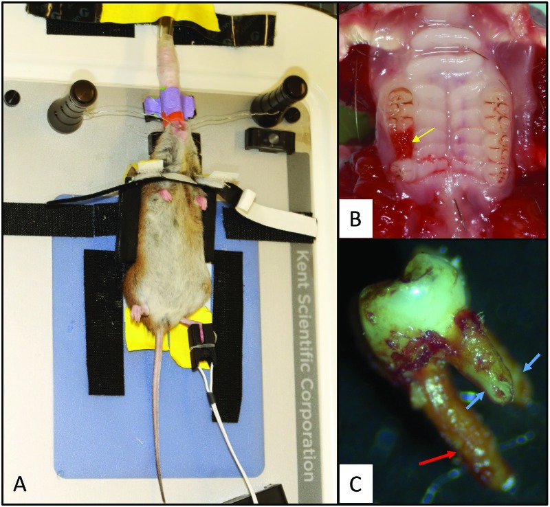
Figure 4.
Tooth extraction procedure in rice rats. (A) Rice rats were placed on a heated surgical platform in dorsal recumbency and stabilized by using a custom-made support device. The head was stabilized by placing surgical wire affixed to 2 magnetic stabilizers caudal to the maxillary incisors. Isoflurane was administered to the rats through a nasal cone. A pulsimeter was placed on the foot to measure SpO2 and heart rate. (B) Dorsal aspect of the palate; the yellow arrow indicates the extraction socket after removal of the right second maxillary molar. (C) A complete extracted second molar, showing the larger, palatal root (red arrow) and 2 smaller, mesial and distal buccal roots (blue arrows). All 3 roots were intact after extraction in 80% of the rats, as depicted in the photo, whereas in 20% of rats, a small apical piece of one of the small buccal roots was broken.

