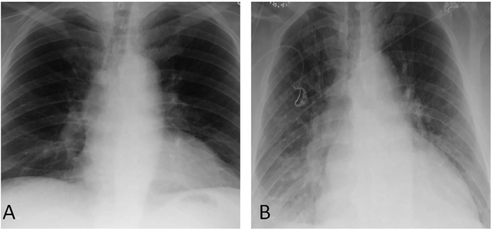
FIGURE 2.
Last available posteroanterior (PA) chest film obtained 6 years prior to admission notes normal cardiac size and no evidence of pulmonary infiltrates (A). PA chest x-ray upon admission (B) notes interval development of diffuse hazy interstitial opacities predominately at the base, cardiomegaly, and pulmonary venous hypertension

