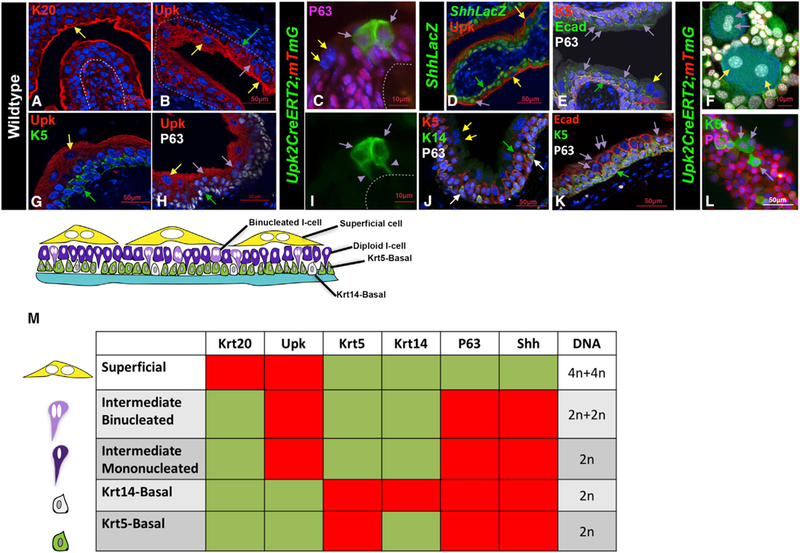
Figure 1. Identification of a New Binucleated Intermediate Cell Population Likely to Be Direct Superficial Progenitors.

(A) Immunofluorescence staining shows K20 expression in sections of bladder from wild-type adult mice. The yellow arrow points to a K20-positive S-cell. Scale bar, 50 µm.
(B) Immunofluorescence staining for Upk3 in a section of bladder from a wild-type adult mouse. Scale bar, 50 µm.
(C) A cryosection from the urothelium of a Upk2CreERT2;mTmG mouse 10 days after tamoxifen treatment shows expression of membrane-bound Gfp. The purple arrows point to Gfp-labeled mononucleated I-cells that form very close to one another, connected to the basement membrane by thin cytoplasmic extensions (purple arrowheads in I). Scale bar, 10 µm.
(D) Immunofluorescence staining for LacZ, P63, and Upk in sections of a bladder from an adult ShhLacZ reporter mouse (Harfe et al., 2004). The yellow arrow points to a LacZ-negative S-cell. Purple arrows point to LacZ-positive intermediate cells, and the green arrow points to a LacZ-positive basal cell. Scale bar, 50 µm.
(E) Immunostained paraffin section from an adult wild-type mouse shows expression of K5, E-cad, and p63. The yellow arrows points to a binucleated S-cell. The double purple arrows points to binucleated I-cells. The green arrow points to a basal cell. Scale bar, 50 µm.
(F) Urothelial cells isolated from a tamoxifen-induced adult Upk2CreERT2;mTmG mouse. Scale bar, 10 µm.
(G) An immunostained paraffin section from a wild-type mouse shows expression of Upk and K5. The yellow arrow points to an S-cell, and the green arrow points to the K5-labeled basal cell. Scale bar, 50 µm.
(H) An immunostained paraffin section from the urothelium of an adult mouse showing expression of Upk and p63. The yellow arrow points to an S-cell; the purple arrows point to intermediate cells; and the green arrow points to a basal cell. Scale bar, 50 µm.
(I) A cryosection from the urothelium of a Upk2CreERT2;mTmG mouse 10 days after tamoxifen treatment shows expression of membrane-bound Gfp. The purple arrows point to Gfp-labeled mononucleated I-cells that form very close to one another and are connected to the basement membrane by thin cytoplasmic extensions. Purple arrowheads denote the Gfp+ cytoplasmic extensions connecting the I-cell to the basement membrane. Scale bar, 10 µm.
(J) Immunofluorescence staining for K5, K14, and p63 in sections of bladder from a wild-type adult mouse. The white arrows point to K14+ basal cells. Scale bar, 50 µm.
(K) A paraffin section from an adult mouse stained with Ecad, K5, and P63. Double purple arrowheads denote binucleated I-cells. Single purple arrow denotes a mononucleated I-cell. Scale bar, 50 µm.
(L) Cells washed from a Upk2CreERT2;mTmG adult mouse urothelium stained with K5 and P63. Scale bar, 50 µm.
(M) Combinatorial markers used to distinguish different urothelial populations.
