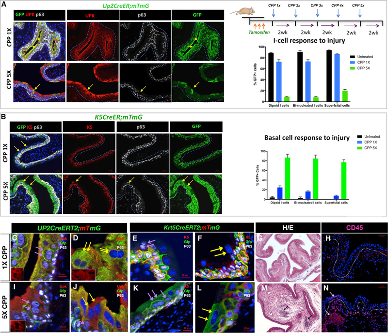
Figure 5. S-Cells Are Generated from Either I or Basal Cells during Regeneration, Depending on the Extent of Injury.

(A) Paraffin sections of bladders isolated from Up2CreERT2;mTmG mice after one or five rounds of CPP-induced injury and regeneration. The yellow arrows point to lineage-marked S-cells. Sections were stained for expression of Gfp, Upk, and P63. Scale bar, 50 µm. The right-top panel shows a schematic description of the injury model. The graph shows the distribution of Gfp-expression in mononucleated I-cells, binucleated I-cells, and S-cells in Up2CreERT2;mTmG that were untreated, treated 13 with CPP, and after 53 CPP treatment.
(B) Paraffin sections from K5CreER;mTmG after one or five rounds of CPP-induced injury and regeneration. Sections were stained for expression of Gfp, K5, and p63. Scale bar, 50 µm. The graph in the right panel shows the distribution of Gfp-expression in mononucleated I-cells, binucleated I-cells, and S-cells in untreated K5CreER;mTmG mice and in K5CreER;mTmG mice after 13 CPP and 53 CPP treatment. Scale bar, 50 µm.
(C) A section from a Up2CreERT2;mTmG mouse treated 13 with CPP showing lineage-marked binucleated I cells (purple arrows). Inset shows Upk expression in a low power magnification of the same section.
(D) A section from a Up2CreERT2;mTmG mouse treated 13 with CPP showing lineage-marked S-cells (yellow arrows). Inset is Upk staining alone in a lower magnification image of the same section.
(E) A section from a Krt5CreERT2mTmG mouse after 1 round of CPP showing lineage-marked basal cells as well as a binucleated I cell that is unlabeled (purple arrows).
(F) A section from a Krt5CreERT2mTmG mouse after 1 round of CPP showing Gfp-labeled basal cells as well as a binucleated S cell that is unlabeled (yellow arrows).
(G) Representative H&E-stained section from the bladder of a mouse treated with a single dose of CPP. Scale bar, 50 µm.
(H) Expression of CD45 in sections from bladders of mice treated with a single dose of CPP. Scale bar, 50 µm.
(I) A section from a Upk2CreERT2mTmG mouse showing lack of Gfp-labeling in binucleated I cells after five cycles of CPP-induced injury and repair (purple arrows). Inset is Upk staining alone in a lower magnification image of the same section.
(J) A section from a Upk2CreERT2mTmG mouse after 5X CPP treatment. Yellow arrows point to b inucleated S-cells, which are unlabeled.
(K) A section from Krt5CreERT2mTmG mouse showing Gfp-labeled binucleated I-cells (purple arrows) after five cycles of CPP-induced regeneration and repair.
(L) A section from Krt5CreERT2mTmG mouse showing Gfp-labeled binucleated S-cells (yellow arrows) after five cycles of CPP-induced regeneration and repair.
(M) Representative H&E-stained section of a bladder from a mouse treated with five doses of CPP. Scale bar, 50 µm.
(N) Expression of CD45 in sections from a mouse treated with five doses of CPP. Scale bar, 50 µm.
