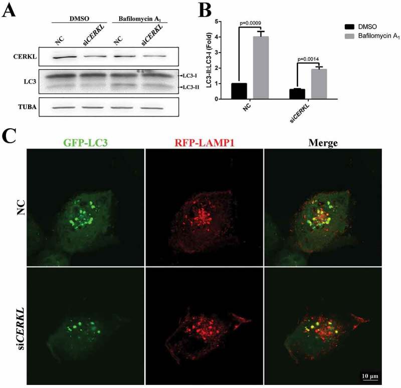
Figure 3.

Autophagic flux was impaired in CERKL-depleted ARPE-19 cells. (a and b) Immunoblotting and quantification of the levels of endogenous LC3-II:LC3-I ratio in negative control (NC) and CERKL-depleted ARPE-19 cells under DMSO and 2.5 nM bafilomycin A1 condition for 3 h. Means± SEM of 5 repeats are shown. (c) Immunostaining analysis of the colocalization GFP-LC3 and RFP-LAMP1 in NC and CERKL-depleted ARPE-19 cells. Scale bars: 10 μm.
