Figure 1.

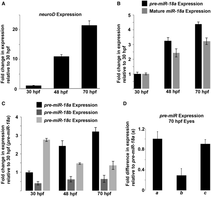
During development, pre‐miR‐18a expression increases proportionally along with neuroD mRNA and mature miR‐18a. (A) neuroD mRNA expression in the developing brain and retina between 30 and 70 hpf. (B) Fold changes in the expression of pre‐miR‐18a and mature miR‐18a in the developing brain and retina between 30 and 70 hpf. (C) Fold changes in the expression of pre‐miR‐18a, pre‐miR‐18b, and pre‐miR‐18c in the developing brain and retina between 30 and 70 hpf. (D) Fold difference in the expression between pre‐miR‐18a (a), pre‐miR‐18b (b), and pre‐miR‐18c (c) in the eyes only at 70 hpf. Error bars represent standard deviation; single biological replicates were used per time point, n = 40 whole heads (A–C) or 40 whole eyes (D) from the AB WT embryos per sample.
