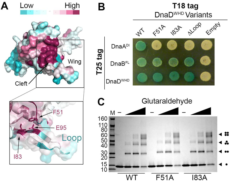
Figure 3. The DnaDWHD Wing Forms a Binding Cleft for DnaA.
(A) The crystal structure of DnaDWHD (PDB 2V79) is shown as a surface representation and colored according to sequence conservation as indicated in the legend (top). The boxed inset shows a semi-transparent surface with F51, I83 and E95 represented as sticks. (B) B2H of T18-tagged DnaDWHD variants co-expressed with either T25-tagged DnaADI, DnaBFL or DnaDWHD. (C) SDS-polyacrylamide gel stained with coomassie blue showing the glutaraldehyde crosslinking of wild type DnaDWHD (WT) or the F51A and I83A variants to reveal self-interactions. The various oligomeric forms are symbolized by dots at the right-hand side of the gel, with each dot representing one DnaDWHD protomer. The molecular weight marker is labeled on the left-hand side in kDa.

