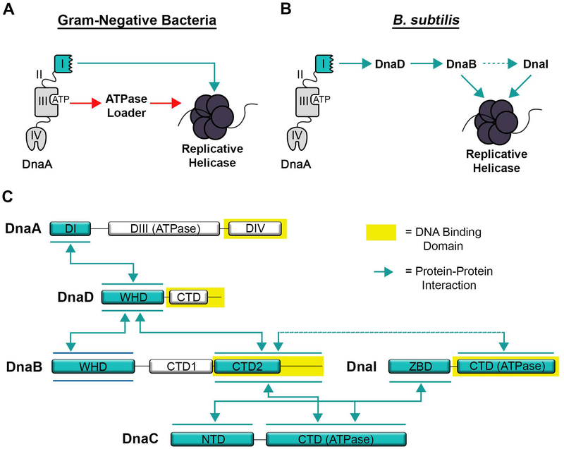
Figure 7. Model of Protein and DNA Interacting Domains Used for Initiation.
(A) Schematic of DnaA interactions from Gram-negative bacterial systems. A direct interaction between DnaA and the replicative helicase representing the E. coli system is shown with a blue arrow, while the interaction between DnaA and the ATPase loader representing A. aeolicus is shown with a red arrow. (B) Schematic of interactions involved in initiation for B. subtilis with interacting proteins connected by blue arrows. (C) Schematic of interacting domains for the initiation proteins in B. subtilis. Double-headed arrows connect domains that interact with each other. DNA binding domains are highlighted in yellow. Note that the order of interaction between DnaB, DnaI and the DnaC helicase are not known. The line connecting DnaB and DnaI is dotted to represent a weak interaction.

