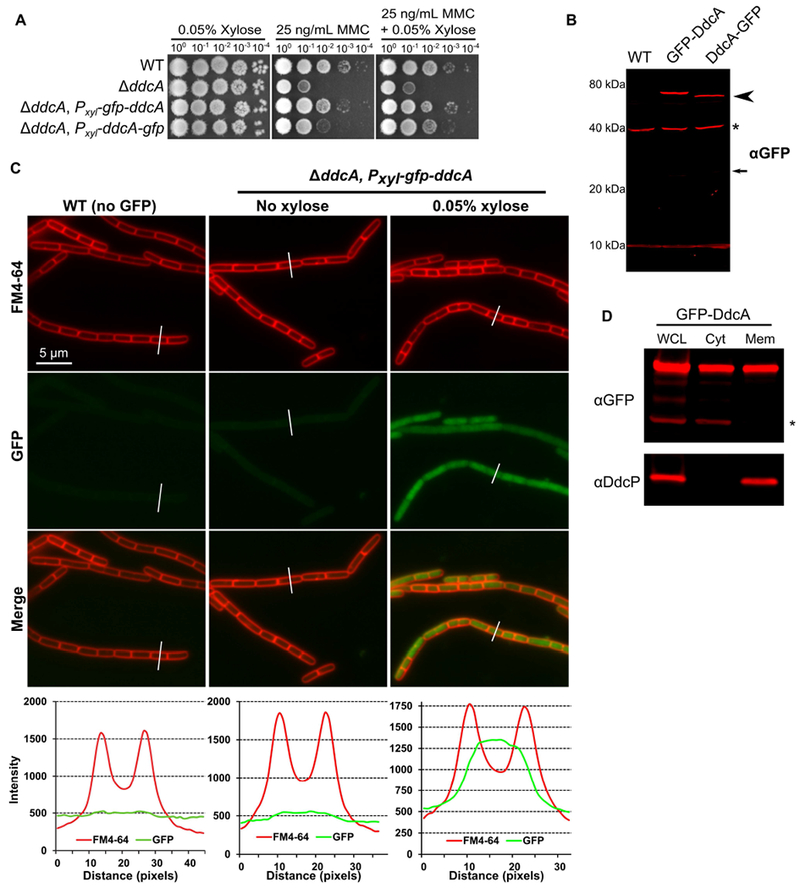
Figure 6. GFP-DdcA is an intracellular protein and is present in the cytosolic and membrane fractions.

(A) Spot titer assay using B. subtilis strains WT (PY79), ΔddcA (PEB357), ΔddcA amyE∷Pxyl-gfp-ddcA (PEB854), and ΔddcA amyE∷Pxyl-ddcA-gfp (PEB856) spotted on the indicated media. (B) Western blot of cell extracts from B. subtilis strains WT (PY79), ΔddcA amyE∷Pxyl-gfp-ddcA (PEB854), and ΔddcA amyE∷Pxyl-ddcA-gfp (PEB856) using antiserum against GFP. The arrowhead highlights the slightly increased mobility of DdcA-GFP, and the asterisk denotes a cross-reacting species detected by the GFP antiserum. The smaller arrow indicates the expected migration of free GFP. (C) Micrographs from WT (PY79) and ΔddcA amyE∷Pxyl-gfp-ddcA (PEB854) cultures grown in S750 minimal media containing 1% arabinose with (far left and right panels) or without (middle panels) 0.05% xylose. Images in red are the membrane stain FM4-64, green are GFP fluorescence and the bottom images are a merge of FM4-64 and GFP fluorescence. The white lines through cells in the images are a representation of the line scans of fluorescence intensity generated in ImageJ and plotted below the micrographs. Scale bar is 5 μm. (D) Western blot of whole cell lysate (WCL), cytosolic fraction (Cyt), and membrane fraction (Mem) from ΔddcA amyE∷Pxyl-gfp-ddcA (PEB854) cell extracts using antisera against GFP (upper panel) or DdcP (lower panel). The asterisk denotes a cross-reacting species detected by the GFP antiserum.
