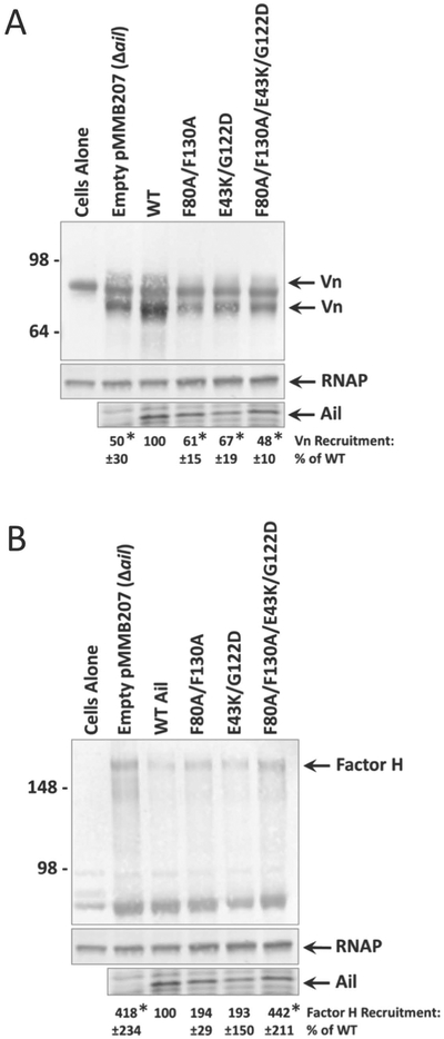
Figure 5. Loss of Ail-mediated recruitment of complement regulatory factors at lower bacterial concentration.
Overnight cultures grown in the presence of 100μM IPTG (to induce Ail expression) were mixed with 50% NHS to a final OD620 = 0.25. Mixtures were shaken vigorously at 37°C for 30 minutes. Samples were centrifuged and cell pellets were washed, then subjected to Western blotting for complement regulatory factors: A) vitronectin (Vn) and B) factor H. Western blots are accompanied by Coomassie-stained gel showing Ail expression in the same samples, as well as the loading control anti-E. coli RNA polymerase alpha. Molecular weight markers are indicated on the left. The cells alone lane represents Y. pestis in the absence of NHS. Quantification of band intensity was performed using at least 3 independent experiments with ImageJ software (NIH). Intensity of bands corresponding to complement regulator recruitment is shown as a percentage of wild-type Ail-mediated recruitment (normalized to 100%) in each individual blot. Significance was determined using one-way ANOVA with Tukey’s post hoc test. *, p-value < 0.05 when compared to a strain expressing wild-type Ail.

