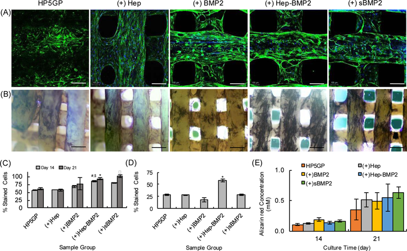
Figure 8.

A) Confocal images of hMSCs cultured on 3D printed scaffolds for 14 days in osteogenic differentiation media, and immunostained for human osteocalcin (OC) (green). Cell nuclei are stained with DAPI (blue). Scale bars are 200 μm. B) Brightfield images of hMSCs stained for alkaline phosphatase (ALP, dark blue/purple) after 14 days of culture in osteogenic media. Scale bars are 400 μm. C) Percentage of cells stained positive for OC corresponding to (A). For day 14, #p<0.2 for Hep-BMP2 as compared to HP5GP and (+)Hep, $p<0.5 for Hep-BMP2 as compared to (+)BMP2. For day 21, αp<0.02 sBMP2 group as compared to HP5GP, (+)Hep, and (+)BMP2, and *p<0.4 for (+)Hep-BMP2 as compared to HP5GP and (+)Hep. D) Percentage of cells stained positive for ALP corresponding to (B). *p<0.001 for (+)Hep-BMP2 as compared to other sample groups (n=3). E) Alizarin Red (AR) staining quantification results using fluorometric analysis depicting AR concentration (mM) for each scaffold after 14 and 21 days of culture in osteogenic induction media.
