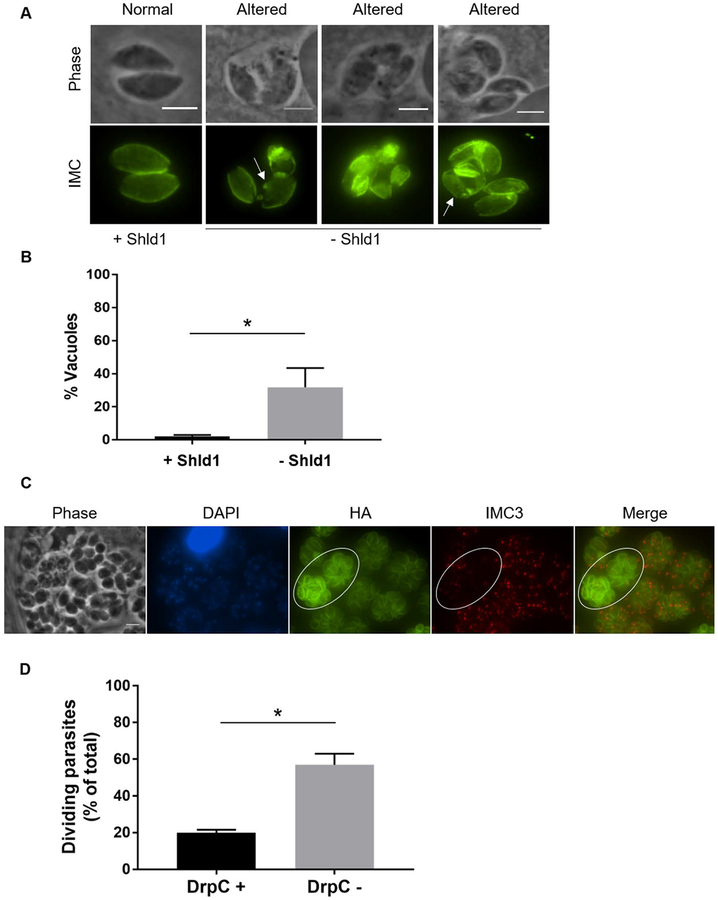
Figure 7. Inner membrane complex formation and parasite division in absence of TgDrpC.
A) Immunofluorescence analysis of DrpC-HA-DD parasites grown for 42 hours in absence of Shld1. Anti-IMC3 antibody was used to detect the inner membrane complex. Images of parasites with either normal or altered IMC are shown. Arrows indicate IMC alterations. Scale bar = 3 μm. B) Percentage of vacuoles with altered IMC structure at 42 hours with or without Shld1. (n=3, ±SD) (*p<0.001). C) Intracellular DrpC-HA-DD expressing parasites grown for 42 hours without Shld1 were stained with antibodies against HA to detect TgDrpC (red). To detect dividing parasites, samples were co-stained with either DAPI (blue) and IMC3 antibodies (green). Circled area indicates dividing parasites. Scale bar = 6 μm. D) Percentage of vacuoles with or without DrpC signal in which parasites were dividing at 42 hours without Shld1. (n=3, ±SD) (*p<0.001).

