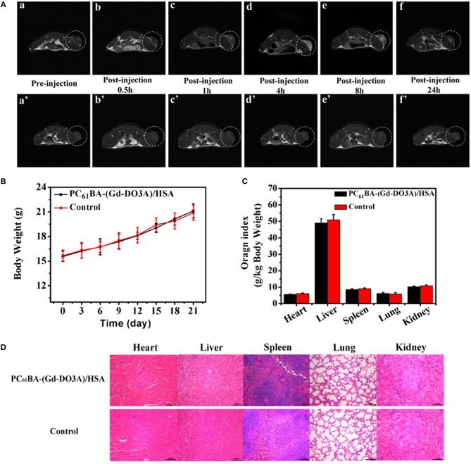
Figure 5.
(A) T1-weighted MRIs of tumor-bearing BALB/C mice before (a) and after injecting PC61BA-(Gd-DO3A)/HSA (0.04 mmol Gd3+/kg bw) at 0.5 (b), 1 (c), 4 (d), 8 (e), and 24 h (f) and those of tumor-bearing BALB/C mice before injection (a′) and after injection of Gd-DO3A (0.04 mmol Gd3+/kg bw) at 0.5 (b′), 1 (c′), 4 (d′), 8 (e′), and 24 h (f′). The tumor tissue is painted in the white dotted ring. (B) Changes in the bw of mice which were injected with the agent or saline (C) Evaluation of organ directories of the mice treated with both presented agent and saline as control sample. (D) histologiacl studies of mice injected with the suggested agent (above) and saline (bottom) as control. Adopted from Zhang et al. (2016) with the Elsevier Permission.

