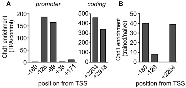
Figure 6.

Chd1 accumulates at the Egr1 gene locus upon stimulation. (A) Egr1 expression was induced with 12-O-tetradecanoylphorbol-13-acetate (TPA) in MLP29 cells, and chromatin immunoprecipitation (ChIP) analysis was performed with an antibody against Chd1 before and at 30 min after induction. (B) Hippocampi were isolated from naïve C57BL/6N mice and from mice 20 min after completion of the training session in the OLM paradigm, and ChIP analysis was performed on chromatin pools from HC of four mice per group. Antibody-bound DNA was detected by qPCR with primers spanning the indicated positions at the Egr1 genomic locus (numbering relative to the transcriptional start site, TSS). Enrichment of signals from TPA induced vs. non-induced conditions (A) and naïve vs. trained animals (B), respectively, is shown for amplicons located in the promoter and coding region, respectively.
