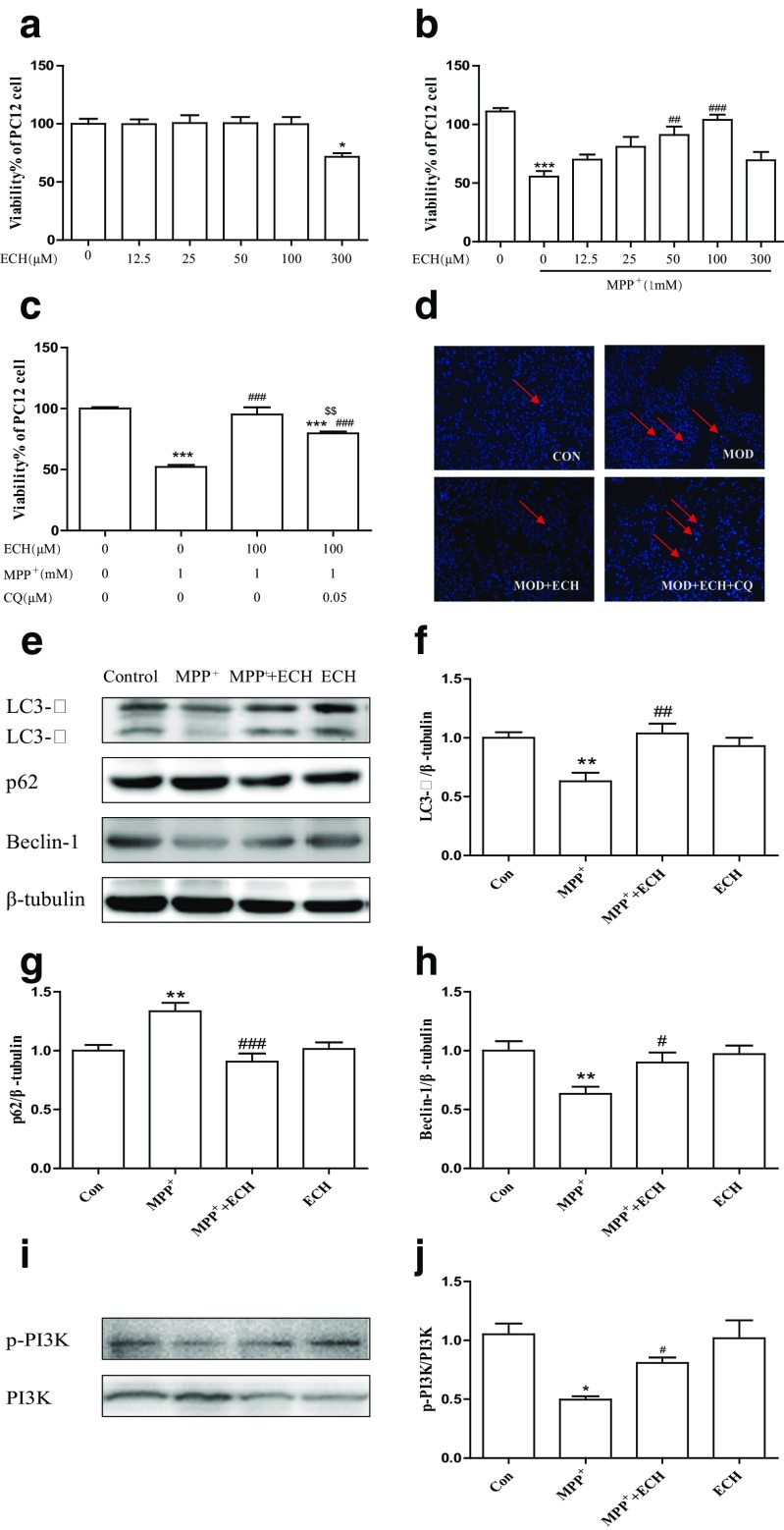
Fig. 4.
ECH protected the viability of MPP+ induced PC12 cells by increasing autophagy Protective effect of ECH on viability of PC12 cells injured by MPP+. Cell viability was determined by MTT assay. Data are expressed as percentage of the viability of cells, control taken as 100% viability. a Effect of different concentration of ECH on cell viability in PC12 cells (determine the non-toxic dosages of ECH). PC12 cells were incubated with ECH (12.5–300 μM) for 24 h. b Effect of ECH on cell viability changes in MPP+-induced PC12 cells. PC12 cells were pre-incubated with different concentration of ECH (0, 25, 50, 100 μM) for 1 h. Then, MPP+ was added to the wells at a final concentration of 1 mM and incubated for another 24 h at 37 °C. c Protective effect of ECH on cell viability was reduced by CQ. Values are presented as means ± standard error (n = 6). *p < 0.05, **p < 0.01, ***p < 0.001, compared with the con group; #p < 0.05, ##p < 0.01, ###p < 0.001, compared with the MPP+ group. d Apoptotic cells were examined in terms of changes in cell morphology by Hoechst 33342 staining (red arrows were represented as apoptotic cells). e‑j Western blotting analysis and quantification of relative LC3, p62, Beclin1, p-PI3K and PI3K protein abundance in the PC12. Tubulin protein served as the internal control. Values are presented as means ± standard error (n = 3). *p < 0.05, **p < 0.01, compared with the con group; #p < 0.05, ##p < 0.01, ###p < 0.001, compared with the MPP+ group

