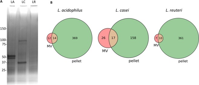
Figure 3.
Protein composition of Lactobacillus MVs. (A) Gel-Code Blue-stained SDS-PAGE of purified L. acidophilus (LA), L. casei (LC), and L. reuteri (LR). MVs with equal number of MVs loaded. For the sake of clarity the Escherichia coli OMVs that were run in parallel were removed from this image. A complete gel image can be found in the Supplemental Material. (B) Venn diagrams of the identified proteins that are unique or in common between the MVs and pellets of each Lactobacillus species.

