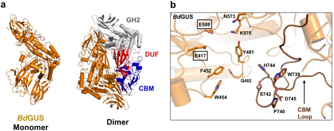
Figure 3.
Crystal structure of NL BdGUS. (a) Monomeric tertiary structure and dimeric quaternary structure of BdGUS (orange). A single monomer is highlighted in orange and the identical monomer is shown with the glycosyl hydrolase 2 (GH2) in white, and the C-terminal domains (domain of unknown function, DUF; carbohydrate binding motif; CBM) in red and blue, respectively. (b) Active site of BdGUS with the CBM loop indicated by an arrow. Catalytic glutamates are boxed.

