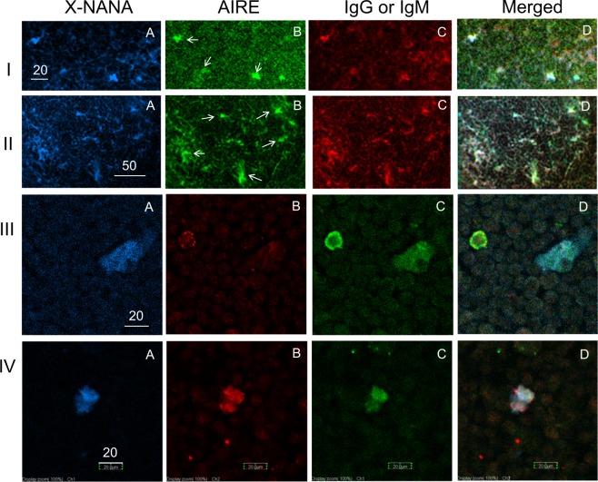
Figure 2.
Expression of AIRE in Neu-medullocytes. (I and II); thymus sections from AKR mice (male, 12 W) were used. (A) Stained with X-NANA; (B) stained with anti-AIRE (anti-AIRE and FITC-conjugated goat anti-rabbit IgG); (C) stained with R.R.-F(ab′)2-anti-mouse IgG (I) or R.R.-F(ab′)2-anti-mouse IgM (II); (D) merged (A–C). The AIRE-positive cells that are indicated by arrows in B are X-NANA and IgG or IgM triple-positive cells (white) in D. (III and IV); These cells were prepared from three thymuses from C57BL/6 mice (male, 6 W) by enzyme digestion and separated into the total thymocyte (T fraction) (III) and E (E1 + E2) fractions (IV). (A) stained with X-NANA; (B) stained with anti-AIRE coupled with rhodamine (TRITC)-conjugated donkey anti-rabbit IgG); (C) stained with FITC-F(ab)2-donkey anti-mouse IgG; (D) merged (A–C). Scale bars are 20, 50, 20 and 20 μm for (I, II, III and IV), respectively.

