
Figure 4.
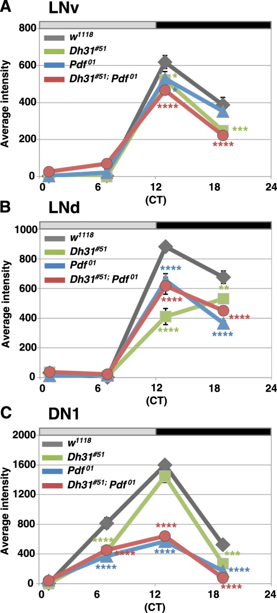
Dh31 mutation did not enhance the abnormal molecular oscillations caused by Pdf mutation. The average levels of VRI expression in 10 brain hemispheres in each subgroup of clock cells (LNv, LNd and DN1) among WT, Dh31#51, Pdf01 and Dh31#51;Pdf01 double mutants at the indicated time points ((circadian time (CT) 1, 7, 13 and 19) in DD3. LNv (A), LNd (B) and DN1 (C). The detailed data of the average intensity are shown in Table S5 The variation of the average intensity in a day in each genotype was compared using two-way ANOVA and Sidak’s multiple-comparison test. The results for comparisons with WT flies at each time point are shown: ****P < 0.0001, ***P < 0.001, **P < 0.01 or *P < 0.05. The remaining comparisons are shown in Table S4.