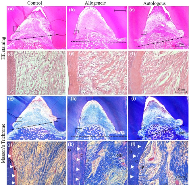
Figure 4.
Histological analysis of periodontal regeneration by allogeneic ADMPC. Representative sections of control and experimental samples were examined by hematoxylin-eosin and Masson’s trichrome staining. The dotted line denotes the base of the defect. Minimal new bone regeneration was observed in the (a) control sample, whereas a greater amount of new bone regeneration was found in the (b) allogeneic and (c) autologous ADMPC transplants. (d–f) Higher magnified sections of the framed regions in (a–c), indicating immune cell infiltration and collagen fiber formation. (g–i) Low magnification of MT-stained images of the control and experimental samples observed for cementum and periodontal tissue regeneration from above the notch of the defect area, as indicated by black squares in the photographs. (j) High magnification sections of the framed region in (g) showing the original cementum area replaced with epithelial tissue (E, arrowhead). (k, l) High magnification sections of the framed region in (h and i) showing new cementum (C, arrowhead) and periodontal ligament (P) fiber formation. E, epithelial tissue; C, cementum; P, periodontal ligament; NB, New bone.

