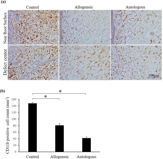
Figure 5.

Immunohistochemical analysis of the defect area with anti-CD11b-antibody. Immunohistochemical images showing CD11b stained cells in control, allogeneic and autologous samples. CD11b positive cells were shown as dark brown color. (a) Photomicrographs adjacent to cementum (upper panel) and central part of the furcation defect area (lower panel) showed high expression of CD11b-stained cells in the control sample, but weak expression was observed in the allogeneic and autologous ADMPC transplants. (b) Quantitative analysis of CD11b-stained cells demonstrated significantly high immune cell infiltration in the control sample compared with the allogeneic and autologous groups. Data are expressed as the mean ± S.D. *p < 0.05 compared with the control.
