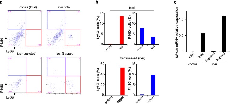
Figure 3.
Elimination of Mincle mRNA by depletion of neutrophils and macrophages/monocytes from injured SN cells. (a) Representative flow cytometry dot plots of Ly6G+ and/or F4/80+ cells and (b) the proportions of these cells in samples of contralateral total, ipsilateral total, ipsilateral depleted fraction, and ipsilateral trapped fraction cells 12 h after PNI. (a) Lower-right (red frame) and upper-left (blue frame) quadrant of the panels representing neutrophils (Ly6G+) and macrophages/monocytes (F4/80+), respectively. Note that neutrophils and macrophages/monocytes were adequately removed from the ipsilateral depleted fraction (lower-left panel in a and lower panels in b). (c) The Mincle mRNA expression in each sample. The expression level of Mincle gene was normalized to that of Gapdh gene. PCR amplifications were done in triplicate. Data are representative of two independent experiments. Note that Mincle mRNA expression in the ipsilateral depleted fraction was virtually eliminated.

