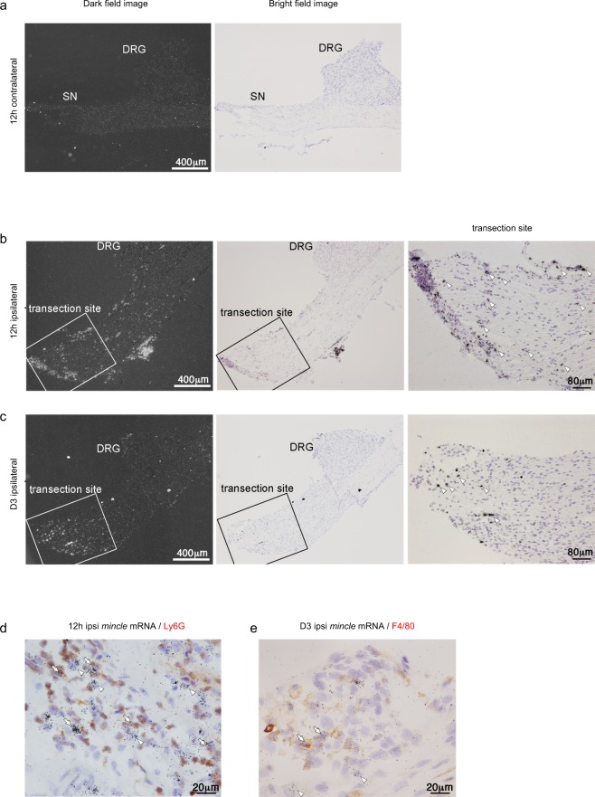
Figure 4.
PNI up-regulates Mincle mRNA at the transection site. (a–c) Dark field and bright field images of the same area of ISHH revealed mRNA distribution of Mincle mRNA after PNI in the L4 DRG and SN, 12 h after PNI, contralateral (a), ipsilateral (b), and 3 days after PNI, ipsilateral site (c). Arrowheads indicate positive cells. Scale bar: Dark field and bright field images, 400 μm, transection site, 80 μm. Counterstained with haematoxylin. (d,e) Bright-field photomicrographs of combined ISHH for Mincle mRNA with immunostaining with Ly6G or F4/80 in the ipsilateral SN. Arrowheads indicate cells single-labelled with ISHH (aggregation of grains). Arrows indicate cells double-labelled with ISHH (aggregation of grains) and IHC (brown staining). Counterstained with haematoxylin. Scale bar: 20 μm.

