Figure 5.
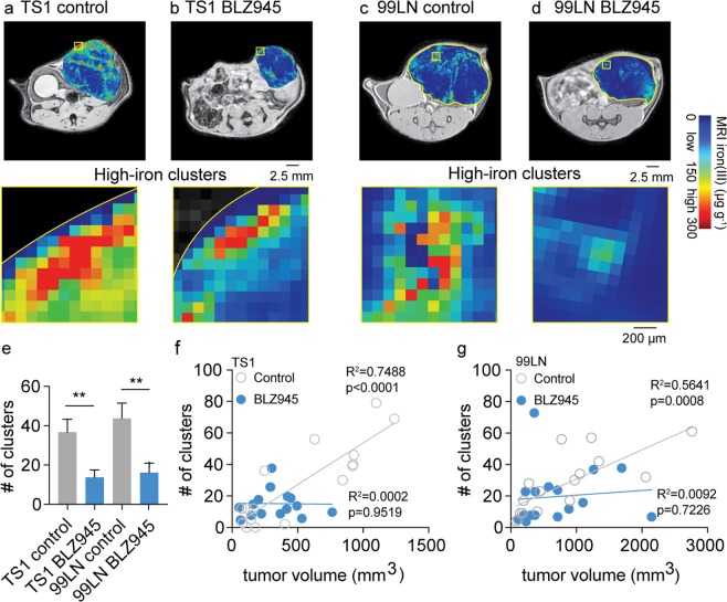
In vivo iron MRI (FeMRI) of murine macrophage iron deposits and correlation between immune and therapeutic CSF1R inhibitor response. Representative in vivo FeMRI axial cross sections of the mammary tumors are shown in control and BLZ945 treated (a,b) TS1, and (c,d) 99LN models. Scale bar 2.5 mm. Expansions show high-iron pixel clusters. Scale bar 200 µm. (e) Number (#) of high-iron FeMRI pixel clusters in the TS1 and 99LN tumors in the CSF1R inhibitor trials (mean + s.e.m. n = 8 mice/group, **p < 0.01 two-tailed unpaired students t-test). Linear correlations between high-iron FeMRI clusters and tumor volumes in the control(ο) and BLZ945-treated(•) (f) TS1 and (g) 99LN MMTV-PyMT tumor models (n = 8 mice/group, R2 and correlation p-value from linear Pearson correlation are shown).

