Figure 7.
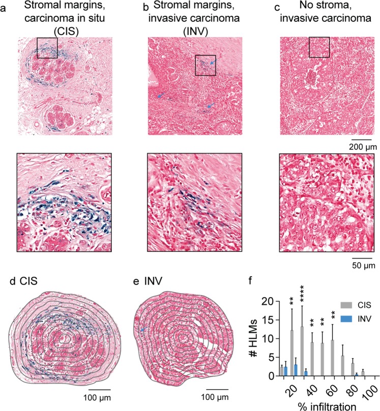
Spatial scores of iron deposits from Prussian blue histology in human breast cancer. Prussian Blue iron histochemistry shows the presence of iron deposits in (a) stromal margins of carcinoma in situ (CIS) and (b) invasive carcinoma (INV, blue arrows). No iron deposits were associated with (c) invasive carcinoma exhibiting poorly defined stromal margins. Scale bar 200 µm. Expansions of boxes in (a–c) shown below. Scale bar 40 µm. Concentric rake region of interest grid overlay used to profile HLMs in (d) CIS and (e) INV fields. Scale bar 100 µm. (f) Iron+ macrophage (HLM) infiltration profiles from Prussian blue histology in CIS and INV fields. (mean + s.e.m. n = 5 fields/cancer subtype, **p < 0.01, ****p < 0.0001, 2-way ANOVA with Sidak’s multiple comparison test).

