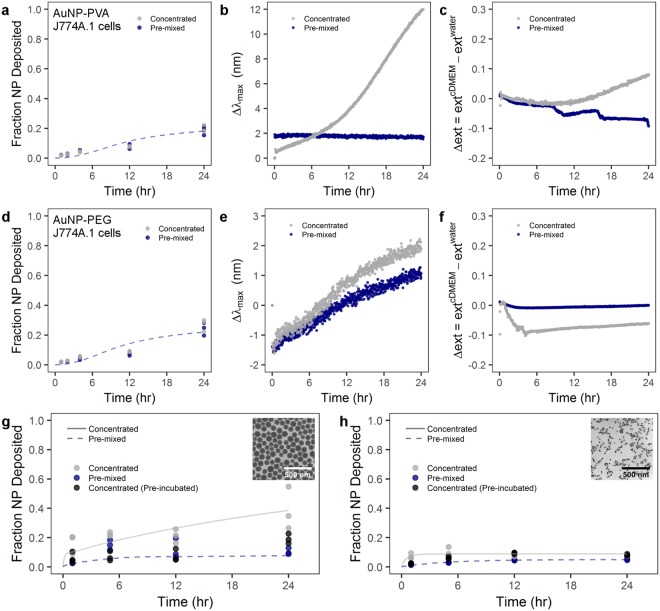
Figure 5.
Administration method effect on particle-cell interaction is heavily dependent on particle material, size, and surface coating. Cellular uptake by J774A.1 murine cells and UV-Vis analysis of (a–c) PVA-coated AuNPs and (d–f) PEG-coated AuNPs. J774A.1 uptake of (g) PVP-coated SiO2 NPs and (h) PVP-coated superparamagnetic iron oxide NPs. (b,e) LSPR band shift for PVA-coated and PEG-coated AuNPs, respectively, following the concentrated or pre-mixed administration. (c,f) Time-dependent change of the extinction intensity (Δext) with respect to the extinction in water at maximum for PVA-coated and PEG-coated AuNP, respectively. Lines indicate modified ISDD fit of data with concentrated or pre-mixed assumptions. Inset TEM micrographs show (g) PVP-coated SiO2 and (h) PVP-coated superparamagnetic iron oxide NPs. Scale bars represent 500 nm.

