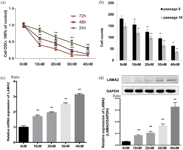
Figure 3.
Demethylation of LAMA2 affects LAMA2 expression and the proliferation of pituitary adenoma cells.
(a) Relative optical density (OD) of GH3 pituitary adenoma cells at 24, 48 and 72 h after treatment with the indicated concentrations of 5-aza-2-deoxycytidine. OD values are reported relative to the control (the concentration of DAC: 0 nmol/l) and were determined using an MTT assay; (b) after 72 h of treatment, the viability of passage two GH3 cells was significantly decreased with increasing concentrations of 5-aza-2-deoxycytidine [passage 8 (black) versus passage 10 (gray)], p < 0.01); (c) RT-qPCR analysis of LAMA2 mRNA expression 72 h after treatment with the indicated concentrations of 5-aza-2-deoxycytidine; (d) levels of the LAMA2 and GAPDH proteins measured in GH3 pituitary adenoma cells at 72 h after treatment with the indicated concentrations of 5-aza-2-deoxycytidine; quantitative analyses of western blotting results in GH3 pituitary adenoma cells are shown.
All data are presented as means ± SEM, *p < 0.05 and **p < 0.01. PCR.
DAC, ; GAPDH, ; GH3, ; LAMA2, laminin subunit alpha 2; mRNA, messenger ribonucleic acid; MTT, ; RT-qPCR, quantitative real-time polymerase chain reaction; SEM, standard error of the mean.

