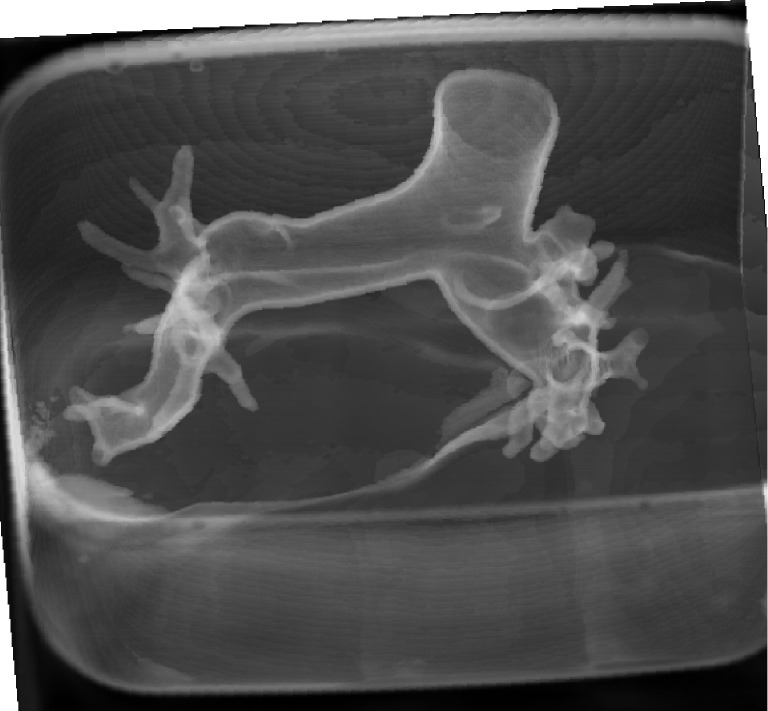
Figure 2.

3D visualization of 3D printed pulmonary artery model which was placed inside the container filled with contrast medium. Since the model was immersed into the water with diluted contrast medium with similar CT attenuation to that of routine CT pulmonary angiography, surface voxel projection was used to create 3D view of the model.
