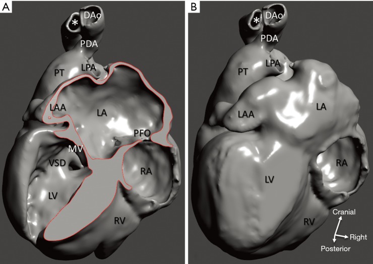
Figure 10.
Interrupted aortic arch and VSD (case 3). (A) 3D model of a cardiac specimen with the left ventricle opened at the pathological examination; (B) the same model with the left ventricle closed by CAD, dorsal view. Orifices of the pulmonary veins are not represented as they were not captured at scanning for technical reasons. Surgical anastomosis between the interposition polytetrafluoroethylene tube (*) and the descending aorta was truncated on the original specimen. DAo, descending aorta; LA, left atrium; LAA, left atrial appendage; LPA, left pulmonary artery; LV, left ventricle; MV, mitral valve orifice; PFO, patent foramen ovale; PDA, patent arterial duct; PT, pulmonary trunk; RA, right atrium; RV, right ventricle; VSD, ventricular septal defect.

