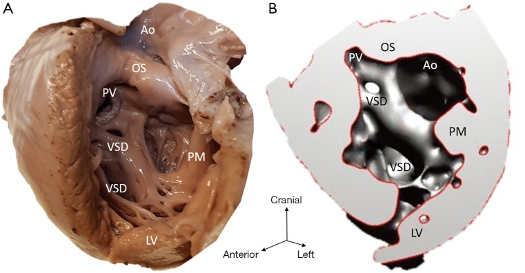
Figure 4.
Multiple VSD (case 4). (A) Cardiac specimen; (B) 3D model of the same heart with the lateral wall of the left ventricle open. Deficiency and misalignment of septal components result in multiple VSDs and appearance that both pulmonary and aortic outlets are connected to the left ventricle. Ao, aortic valvar orifice; LV, left ventricle; OS, outlet septum; PM, papillary muscle; PV, pulmonary valvar orifice; VSD, ventricular septal defect.

