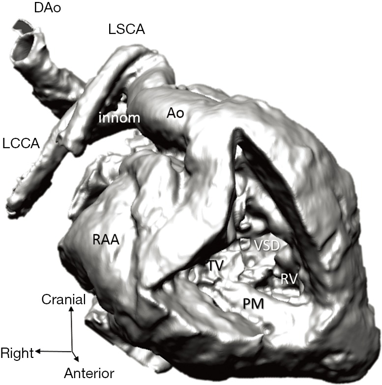
Figure 5.

Representation of the right ventricle and its connexions (3D virtual model of case 1), with transposition of the great arteries and ventricular septal defect. Arch vessels and descending aorta suffered major distortion due to inappropriate positioning of the specimen before scanning. Ao, aorta; DAo, descending aorta; innom, innominate artery; LCCA, left common carotid artery; LSCA, left subclavian artery; PM, papillary muscle; RAA, right atrial appendage; RCCA, right common carotid artery; RV, right ventricle; TV, tricuspid; VSD, ventricular septal defect.
