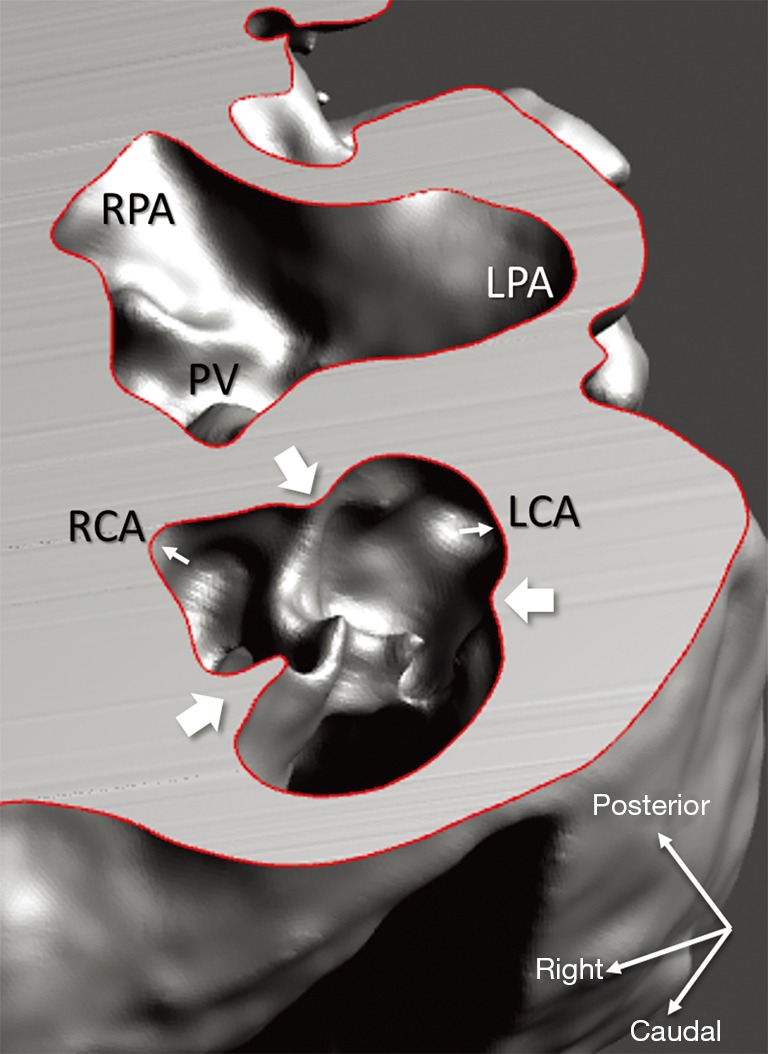
Figure 7.

Representation of the aortic valve (3D virtual model of case 1), with transposition of the great arteries and ventricular septal defect. Anterior aortic root features three sinuses and semilunar valve leaflets hinged by commissures (broad white arrows). Facing sinuses give rise to coronary ostia (narrow white arrows) for the right and left coronary arteries (RCA, LCA). Pulmonary bifurcation lies posterior; incipient segments of the right and left pulmonary arteries (RPA, LPA) are visible as well as the pulmonary valve (PV).
