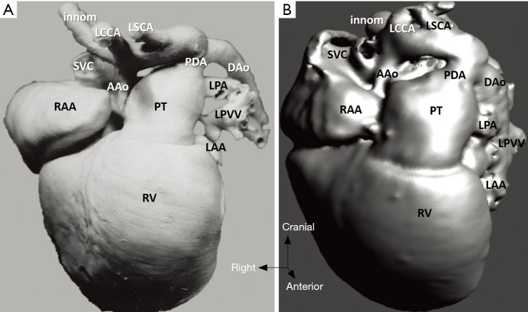
Figure 8.
Hypoplastic left heart syndrome (case 5). (A) Cardiac specimen; (B) 3D model of the same heart, frontal view. The 3D virtual model demonstrates significant accuracy in the segmental anatomy and the arterial structures. AAo, ascending aorta; DAo, descending aorta; innom, innominate artery; LCCA, left common carotid artery; LSCA, left subclavian artery; LAA, left atrial appendage; LPA, left pulmonary artery; LPVV, left pulmonary veins; PDA, patent arterial duct; PT, pulmonary trunk; RAA, right atrial appendage; RV, right ventricle; SVC, superior vena cava.

