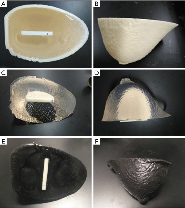
Figure 4.
Photographs of the breast phantom. (A,B) The breast phantom and insert within the breast mold. (C,D) The PVC breast phantom has been removed from the mold and is ready for mammography and MRI. (E,F) An ultrasound breast phantom uses the mixture of PVC, softener and 3.0% graphite power to improve tissue contrast for imaging.

