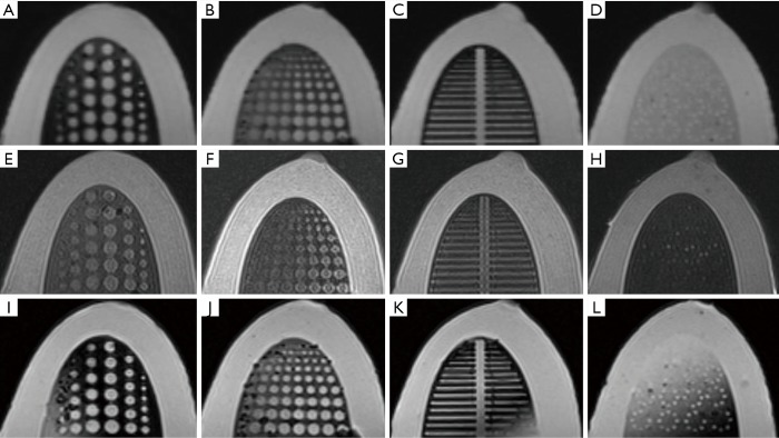
Figure 6.
MRI images of the multi-modal breast phantom with T1WI (A,B,C,D), T2WI (E,F,G,H), and STIR (I,J,K,L) sequences. The first column (A,E,I) shows tumor inserts, the second column (B,F,J) shows depth resolution inserts, the third column (C,G,K) shows fiber inserts, and the forth column (D,H,L) shows microcalcification inserts.

