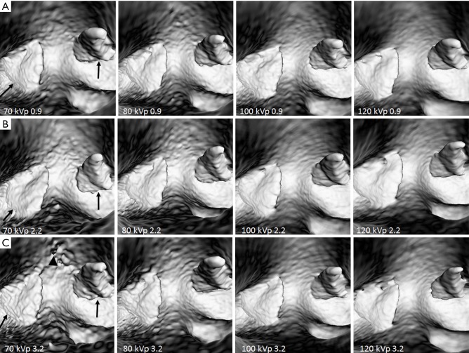
Figure 5.
VIE of thrombus in images acquired with different CTPA protocols. (A,B) Intraluminal views of the thrombus (arrows) are clearly demonstrated with CTPA protocols using different kVp and pitch values of 0.9 and 2.2. (C) when high pitch of 3.2 was used, irregular appearance of the thrombus (arrows) and some artifacts (arrowhead) appeared in the low kVp 70 protocol when compared to other protocols. VIE, virtual intravascular endoscopy; CTPA, computed tomography pulmonary angiography.

