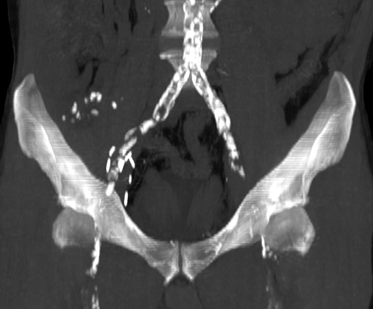
Figure 1.

Coronal MIP image made from an abdominal pelvic CT scan without intravenous iodinated contrast injection. This type of image gives the surgeon a global view of the size and distribution of calcified atheromatous plaques; however, it is not precise enough and does not replace the intraoperative palpation of vessels during kidney transplantation. MIP, maximum intensity projection.
