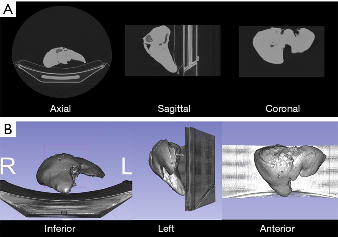
Figure 2.

Computed tomography of 3D printed liver models. (A) Axial, sagittal and coronal views of liver model. Silicone parenchyma has higher attenuation than plastic elements (tumor, vessels); (B) inferior, left and anterior volume rendering views of 3D printed model.
