
Figure 2.
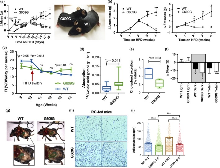
Robust response of progeria mice to high‐fat diet. (a) Male G609G mice (N = 5) and WT (N = 6) controls were switched to a HFD (60% calories from fat) at 11 weeks of age, and cumulative mass change from starting mass was recorded daily. Picture shows mice 4 days after the switch to HFD, a point at which G609G mice experienced a 15% increase of their body weight. (b) The same mice as in (a), switched to HFD, were placed in NMR weekly to determine lean mass and fat mass changes from the initial mass. (c) Food intake (% body weight ingested per day per mouse) in G609G and WT mice fed RC (weeks 9 and 10) and switched to HFD for 4 weeks (weeks 11–14). Mice were single‐housed, and food consumption monitored daily. Graph shows FI weekly average ± SEM (N = 5 male mice per group). (d) G609G mice (N = 10) and WT controls (N = 9) seven weeks of age were administered 14C‐oleic acid via gavage and 3 hr later radioactivity in plasma measured. Aborption calculated as pmol of 14C‐oleic acid per gram (body weight) per hour. Half mice were males and half females. (e) Single‐housed male G609G mice (N = 5) and WT controls (N = 5) were switched to a high cholesterol diet (HCD) at 7 weeks of age. Daily feces were collected twenty days after the switch. Note how the amount of cholesterol excreted in the feces is significantly reduced in G609G mice with respect to WT. (f) Change in percentage sleep immediately after mice were switched to HFD compared to sleep pattern on RC. (g) Necropsy images of WT and G609G mice switched from RC to HFD and maintained on HFD for four weeks. Notice adipose tissue is evident in abdominal cavity of G609G mice, which was not previously present for mice on RC (Figure 1d). (h) Images from histology of H&E stained adipose tissue from WT and G609G mice fed RC. (i) Graph shows quantification of adipocytes size in G609G (N = 9) and WT (N = 10) mice fed RC up to 11 weeks of age, and in mice switched from RC to HFD at 11 weeks of age and maintained in HFD for 4 weeks (N = 5 in each group of both G609G and WT mice)