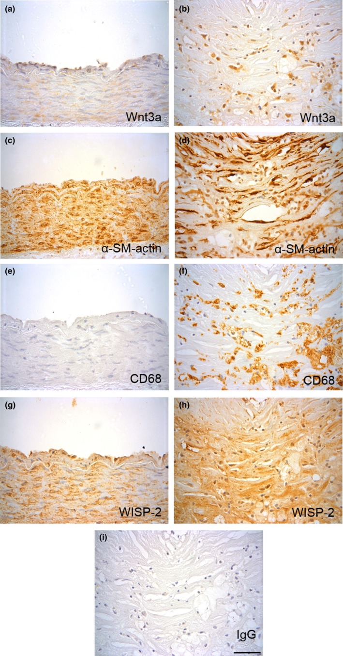
Figure 1.

Wnt3a and WISP‐2 proteins are upregulated in human atherosclerotic plaques. Representative images of immunohistochemistry for Wnt3a (a, b), α‐smooth muscle actin (c, d), CD68 (e, f) and WISP‐2 proteins (g, h) in control (a, c, e & g) and atherosclerotic (b, d, f & h) human coronary arteries. Non‐immune rabbit IgG was included as a negative control for Wnt3a and WISP‐2 antibodies (i). The scale bar represents 50 μm and applies to all images. A lower magnification image of each vessel is available in Supporting Information Figure S1
