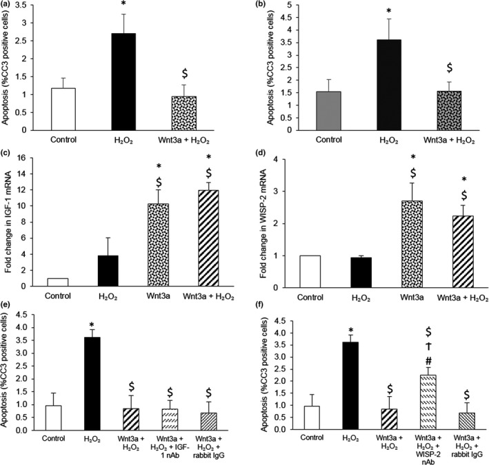
Figure 3.

Wnt3a upregulated multiple pro‐survival genes, but only WISP‐2 was necessary for Wnt3a‐mediated rescue of VSMCs from H2O2‐induced apoptosis. (a, b) Apoptosis was quantified in young TOPGAL mouse VSMCs transfected with either AllStars Negative Control siRNA (a) or WISP‐1 siRNA (b), and then 24 hr later stimulated with 100 μM H2O2, with or without 400 ng/ml recombinant Wnt3a protein, for a further 24 hr. Apoptosis was quantified using CC3 immunofluorescence. The number of CC3 positive cells was counted and expressed as a percentage of the total number of cells viewed. Error bars represent SEM. *p < 0.05 vs. control, $p < 0.05 vs. H2O2, repeated measures ANOVA and Student–Newman–Keuls post hoc test, n = 5. (c, d) IGF‐1 (c) and WISP‐2 (d) mRNAs were quantified by QPCR in young TOPGAL mouse VSMCs stimulated with 100 μM H2O2, with or without 400 ng/ml recombinant Wnt3a protein, for 4 hr. mRNA levels were normalized to 36B4 mRNA levels. Results are shown as the fold change from control. Error bars represent SEM. *p < 0.05 vs. control, $p < 0.05 vs. H2O2, ANOVA and Student–Newman–Keuls post hoc test, n = 3. (e, f) Apoptosis was quantified in young TOPGAL mouse VSMCs stimulated with 100 µM H2O2, with or without 400 ng/ml recombinant Wnt3a protein and 10 µg/ml IGF‐1 neutralizing antibody (nAb) (e) or 10 µg/ml WISP‐2 neutralizing antibody (nAb) (f), for 24 hr using CC3 immunofluorescence. Non‐immune rabbit IgG acted as a negative control. The number of CC3 positive cells was counted and expressed as a percentage of the total number of cells viewed. Error bars represent SEM. *p < 0.05 vs. control, $p < 0.05 vs. H2O2, #p < 0.05 vs. Wnt3a + H2O2, Ϯp < 0.05 vs. Wnt3a + H2O2 + rabbit IgG, repeated measures ANOVA and Student–Newman–Keuls post hoc test, n = 5
