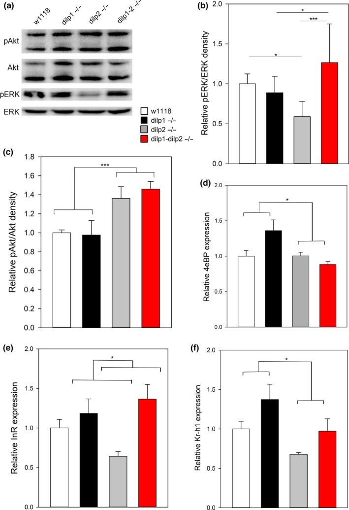
Figure 4.

Components of insulin/IGF and JH signaling regulated by dilp1 and dilp2. (a) pAkt in dissected thorax tissue is increased in dilp2 and double mutants; pERK is decreased in dilp2 mutants in a dilp1‐dependent manner, representative blot. Figures B‐F show significance (*p < 0.05, **p < 0.01) from post hoc pairwise comparisons in two‐way ANOVA. (b) Quantification of thorax pERK/ERK phospho‐westerns, dilp1 × dilp2 interaction p = 0.003, n = 6 per genotype. (c) Quantification of thorax pAkt/Akt phospho‐westerns, dilp1 × dilp2 interaction: not significant, n = 6 per genotype. (d) 4eBP mRNA expression is not elevated in dilp2 mutants but interacts with dilp1, dilp1 × dilp2 interaction p = 0.01, n = 7–9 per genotype. (e) InR mRNA expression is reduced in dilp2 mutants relative to dilp1; dilp2 double mutant p < 0.05, n = 7–9 per genotype. (f) Kr‐h1 mRNA expression is decreased by dilp2 mutation, without significant dilp1 × dilp2 interaction, n = 8–9 per genotype
