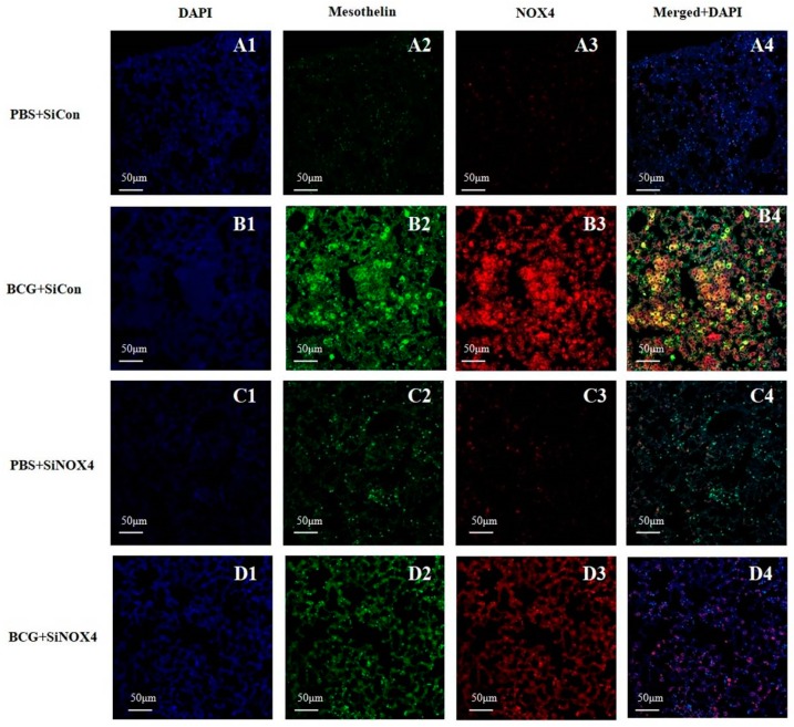
Figure 7.
Activation of NOX4 and mesothelin deposition in a BCG-induced pleurisy mouse model. NOX4 and mesothelin (markers for PMCs) were subjected to immunofluorescence staining. A1–D1: DAPI staining. A2–D2: Immunofluorescence staining of mesothelin (green color). A3–D3: Immunofluorescence staining of NOX4 (red color). A4–D4: Overlays of immunofluorescence staining and DAPI staining, yellow areas represent colocalization of mesothelin and NOX4. DAPI, 4′,6-Diamidino-2-phenylindole dihydrochloride.

